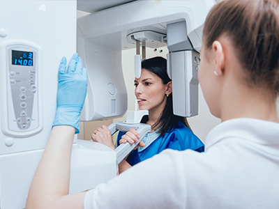

A person receiving a dental examination with a dentist using a magnifying loupe.

Digital Radiography

What modern digital radiography means for your dental care

Digital radiography is the contemporary method dentists use to capture X-ray images using electronic sensors and computer software instead of traditional film. This approach streamlines the entire imaging process: images are created in seconds, displayed on a monitor for immediate review, and stored securely in a digital patient record. Because the workflow is electronic from acquisition to storage, clinicians can move from observation to diagnosis and treatment planning far more quickly than with film.

Beyond speed, digital radiography changes how information flows within the practice and across care teams. High-quality images can be shared instantly with specialists, labs, or other providers when coordination of care is needed. The result is fewer delays, clearer communication, and a smoother patient experience during appointments when imaging plays a role in diagnosis or treatment decisions.

At its core, digital radiography is about giving patients and providers better tools for understanding oral health. The technology reduces manual steps, improves recordkeeping, and supports more precise clinical decisions—advantages that help clinicians deliver consistently higher-quality care.

How digital sensors create clearer, more useful images

Instead of film, digital systems use sensors or plates that convert X-ray energy into electronic signals. Intraoral sensors capture detailed pictures of individual teeth and surrounding structures, while extraoral detectors and scanning plates can be used for broader views of the jaws. The electronic signal is processed by software that enhances contrast and sharpness, making subtle features easier to evaluate without changing the exposure during acquisition.

One of the practical benefits of a digital workflow is the ability to adjust images after they’re taken. Clinicians can zoom, measure, and change contrast to reveal small details—such as early decay between teeth or the margins of a restoration—that might be harder to see on conventional film. These on-screen adjustments preserve the original image while allowing focused review where it matters most.

Digital images are also reproducible and consistent. Because the capture and processing are standardized, clinicians can compare current images with previous ones side-by-side, track changes over time, and document treatment outcomes more reliably. Those consistent records support long-term care planning and help identify trends that inform preventive or restorative decisions.

Patient safety: lowering radiation exposure and improving comfort

One of the principal advantages of digital radiography is its favorable safety profile. Electronic detectors are more sensitive to X-ray photons than film, which means useful diagnostic images can be obtained with a lower dose of radiation. Dentists pair that technological advantage with established safety practices to keep exposure as low as reasonably achievable while still collecting the clinical information needed for diagnosis.

Safety measures are a standard part of the imaging process. Clinicians use the smallest number of images necessary, select exposure settings appropriate for each patient, and employ protective equipment and positioning techniques to minimize unnecessary exposure. These precautions, combined with the efficiency of digital capture, make routine dental imaging both quicker and safer for patients of all ages.

Comfort is another practical benefit. Digital sensors and plates are often designed to be slim and quicker to position than film holders, which can reduce discomfort during intraoral imaging. Because images appear immediately, any need for repositioning is identified and corrected on the spot, reducing the chance of repeated exposures.

Better diagnostics and treatment planning with on-screen tools

High-resolution digital images enhance clinical decision-making. Dentists can detect early stages of tooth decay, assess bone levels, examine root anatomy, and evaluate the fit and integrity of restorations with greater clarity. These capabilities support earlier intervention when problems first appear and help target treatment precisely where it will be most effective.

Digital radiography also supports multidisciplinary care. Images can be exported or examined alongside intraoral photographs, intraoral scans, and clinical notes to create a comprehensive view of a patient’s oral health. When a case requires collaboration—such as implant placement, orthodontic planning, or endodontic treatment—having accurate digital images expedites consultation and helps all providers work from the same reliable data.

Additionally, on-screen tools enable measurements, annotations, and side-by-side comparisons that strengthen documentation. These features are useful for tracking healing after treatment, monitoring the progression of conditions, and explaining findings to patients so they can participate in informed decisions about their care.

What patients can expect during digital X-ray appointments

The digital imaging process is straightforward and usually quick. For intraoral X-rays, a small sensor is placed inside the mouth while the clinician positions the X-ray unit outside the cheek. Extraoral images, such as panoramic views, require brief stillness while the machine rotates around the head. In most cases, the actual exposure lasts only a fraction of a second and the corresponding image appears on the monitor immediately afterward.

Because images are available right away, your clinician can review them with you in real time, pointing out areas of interest and explaining how the findings inform next steps. That immediate feedback helps patients understand the reasons behind recommended treatments and see the visual evidence that supports a diagnosis or monitoring strategy.

All images are retained in the practice’s secure electronic records and handled according to professional privacy standards. Digital storage makes it easy to retrieve past images for comparison at follow-up visits, ensuring continuity of care without the logistical burdens of physical film.

Four Points Family Dental uses digital radiography as part of a commitment to safe, efficient, and patient-centered care. If you have questions about how digital X-rays are used in your treatment or want to learn what to expect at your next visit, please contact us for more information.

Frequently Asked Questions

What is digital radiography?

+

Digital radiography is a modern imaging method that uses electronic sensors and computer software to capture and display dental X-ray images. The system converts X-ray energy into digital signals, producing images almost instantly for clinician review. Because the workflow is digital from capture to storage, images can be integrated directly into a patient’s electronic record for efficient documentation and follow-up.

Digital radiography streamlines diagnosis and treatment planning by making high-quality images available in seconds. Clinicians can adjust contrast, zoom, and measure on-screen to evaluate fine details without re-exposing the patient. This technology supports clearer communication with patients and other providers by enabling straightforward image sharing and side-by-side comparisons.

How does digital radiography differ from traditional film X-rays?

+

Unlike conventional film, digital systems use sensors or plates that capture X-ray information electronically rather than chemically processing film. This eliminates the need for darkroom processing and allows immediate image review, which reduces appointment time and the likelihood of retakes. The electronic capture also enables standardized image processing so clinicians can apply consistent adjustments for improved diagnostic utility.

Digital images are easier to store, retrieve, and compare over time compared with physical film. Because images are reproducible and can be enhanced without altering the original capture, clinicians can document changes in a patient’s oral health more reliably. The ability to export images creates smoother collaboration with specialists, labs, or other providers when multidisciplinary care is needed.

Is digital radiography safe for children and adults?

+

Digital radiography typically requires a lower radiation dose than traditional film imaging because modern electronic detectors are more sensitive to X-ray photons. Dentists follow the principle of keeping exposure as low as reasonably achievable by selecting the minimum number of images needed and using exposure settings appropriate for each patient. Protective measures such as lead aprons and proper positioning are used when indicated to further reduce unnecessary exposure.

For both children and adults, safety practices focus on tailoring the imaging approach to clinical need and patient size. Pediatric imaging protocols often use reduced exposures and smaller sensors to accommodate younger patients. When questions arise about safety, clinicians can explain why each image is recommended and how it contributes to accurate diagnosis or treatment planning.

How does digital radiography improve diagnosis and treatment planning?

+

High-resolution digital images reveal details such as early decay, fine root anatomy, and subtle bone changes that support earlier and more accurate clinical decisions. On-screen tools allow clinicians to measure distances, annotate areas of interest, and compare current images with previous records to track progression or healing. These capabilities help target treatment precisely and reduce uncertainty when planning restorative, endodontic, or surgical procedures.

Digital imaging also facilitates multidisciplinary coordination by enabling clinicians to share consistent, high-quality visuals with specialists and labs. When cases require collaboration for implants, orthodontics, or complex restorations, having reliable digital images expedites consultations and ensures all providers are working from the same information. Clear visual documentation also helps clinicians explain findings to patients, supporting informed decision-making.

What should I expect during a digital X-ray appointment?

+

The imaging process is typically quick and straightforward. For intraoral X-rays a small sensor is placed inside the mouth while the clinician positions the X-ray unit outside the cheek; the exposure itself lasts only a fraction of a second and the image appears immediately on the monitor. For extraoral views, such as panoramic images, the patient remains still for a brief rotation of the machine while the detector captures a broad view of the jaws.

Because images appear right away, your clinician can review them with you during the visit and point out areas of concern or interest. If an image needs repositioning, the team can correct it immediately, which reduces the chance of repeat exposures. All images are then stored securely in the patient record for future comparison and continuity of care.

How are digital images stored and shared securely?

+

Digital images are stored in the practice’s secure electronic records system, where access is controlled according to professional privacy and security standards. Storage in digital format makes retrieval fast and reduces the logistical burdens associated with physical film. Images can also be archived and backed up to protect against data loss while maintaining an auditable record of imaging over time.

When images must be shared with specialists or other providers, clinicians use secure transfer methods that respect patient privacy and comply with applicable regulations. Secure sharing supports coordinated treatment while ensuring that only authorized individuals can access a patient’s diagnostic images and associated clinical notes. Patients can also be shown their images on-screen and provided explanations in real time to support understanding of their care.

Can digital radiography detect dental problems earlier than film?

+

Digital radiography often improves early detection because enhanced contrast and the ability to magnify images make subtle changes easier to identify. Clinicians can adjust image parameters to highlight areas where early decay, small fractures, or bone changes might be developing. These on-screen tools can reveal findings that may be less obvious on traditional film, supporting timely intervention.

Because digital images are reproducible and standardized, dentists can compare current and prior images side-by-side to detect small changes over time. This longitudinal view helps in monitoring conditions and evaluating the effectiveness of treatments. Early detection through clear imaging can reduce the extent of future treatment and support preventive care strategies.

Are digital radiographs used for implant planning, orthodontics, and root canal treatment?

+

Yes, digital radiographs are widely used across many dental specialties because they provide precise views that inform treatment planning and execution. For implant cases clinicians use digital images to assess bone volume and anatomy, aiding in the selection of implant size and placement. In orthodontics panoramic and other extraoral digital images help evaluate growth, tooth position, and skeletal relationships for comprehensive planning.

Endodontic treatment also benefits from high-resolution intraoral images that clarify root canal anatomy, locate additional canals, and document the progression of healing. In each specialty the ability to annotate, measure, and share images enhances collaboration and helps ensure predictable outcomes. Digital imaging supports more accurate diagnostics and a coordinated approach to complex care.

How does Four Points Family Dental minimize radiation exposure during imaging?

+

At Four Points Family Dental clinicians follow established safety protocols to minimize radiation exposure, including using the fewest images necessary for diagnosis and selecting exposure settings appropriate for each patient. Modern digital sensors allow diagnostically useful images at lower doses than film, and staff employ proper positioning and shielding to further reduce risk. Pediatric patients receive additional consideration with size-appropriate settings and techniques.

Regular equipment maintenance and staff training are part of the practice’s approach to safe imaging. Ensuring detectors are calibrated and operators are skilled in positioning reduces the chance of repeats and unnecessary exposure. When patients have concerns about imaging, the team will explain the rationale for each image and answer questions about safety and alternatives.

How do dentists use on-screen tools to explain findings to patients?

+

On-screen tools let clinicians zoom, measure, and annotate images while they review results with patients in real time. This interactive review helps patients visualize issues such as decay, bone loss, or restorative margins and understand why a particular treatment is recommended. Being able to show a clear image and highlight the specific concern improves communication and supports informed consent.

Clinicians can also display side-by-side comparisons with prior images to demonstrate changes or healing over time, which helps patients track progress and engage with preventive strategies. Clear visual explanation combined with plain-language interpretation makes it easier for patients to participate in decisions about their care and to follow recommended treatment plans.

Pay Online

We make paying your dental bill incredibly easy and convenient by offering a secure, dedicated online payment portal. You can quickly access this system from our website using your computer or smartphone at any time, day or night.

Expertise You Can Trust, Care You Can Feel

Experience a higher standard of dentistry for every member of your family. Our doors—and our ears—are always open to your questions. Let us handle the details while you focus on your health. Reach out today to see how we’re redefining the dental visit.

Office Hours

Monday
9:00 am - 4:00 pm
Tuesday
9:00 am - 6:00 pm
Wednesday
9:00 am - 4:00 pm
Thursday
9:00 am - 5:00 pm
Friday
7:30 am - 2:00 pm